Third-Party Research
This contains links and descriptions of research papers written by third-party researchers. These papers have been used by us to back our neural model designs and to give us ideas for future advances and improvements. A lot of these papers are likely to be referenced in our research papers.

Written by Eugene M. Izhikevich in the IEEE Transactions of Neural Networks, Vol. 14, No. 6, November 2003.
A model is presented that reproduces spiking and bursting behavior of known types of cortical neurons. The model combines the biologically plausibility of Hodgkin–Huxley-type dynamics and the computational efficiency of integrate-and-fire neurons. Using this model, one can simulate tens of thousands of spiking cortical neurons in real time (1 ms resolution) using a desktop PC.

Written by Eugene M. Izhikevich and Gerald M Edelman from the Neurosciences Institute on December 27, 2007.
The understanding of the structural and dynamic complexity of mammalian brains is greatly facilitated by computer simulations. We present here a detailed large-scale thalamocortical model based on experimental measures in several mammalian species. The model spans three anatomical scales.

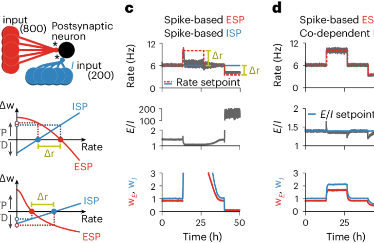
Written by Everton J. Agnes and Tim P. Vogels in Nature Neurosciences 27, 964-974 (2024) on March 20, 2024.
The brain’s functionality is developed and maintained through synaptic plasticity. As synapses undergo plasticity, they also affect each other. The nature of such ‘co-dependency’ is difficult to disentangle experimentally, because multiple synapses must be monitored simultaneously. To help understand the experimentally observed phenomena, we introduce a framework that formalizes synaptic co-dependency between different connection types.

Written by Corey D. Acker, Erika Hoyos, and Leslie M. Loew for eNeuro on May 12, 2016.
EPSPs occur when the neurotransmitter glutamate binds to postsynaptic receptors located on small pleomorphic membrane protrusions called dendritic spines. To transmit the synaptic signal, these potentials must travel through the spine neck and the dendritic tree to reach the soma. Due to their small size, the electrical behavior of spines and their ability to compartmentalize electrical signals has been very difficult to assess experimentally.

Written by Albert Gidon et al. for Science Vol. 367, Issue 6473 on January 3, 2020.
The active electrical properties of dendrites shape neuronal input and output and are fundamental to brain function. However, our knowledge of active dendrites has been almost entirely acquired from studies of rodents. In this work, we investigated the dendrites of layer 2 and 3 (L2/3) pyramidal neurons of the human cerebral cortex ex vivo.

Written by Alexandre Foncelle et al. for Frontiers in Computational Neuroscience Vol. 12 - 2018 on July 2, 2018.
In spike-timing dependent plasticity (STDP) change in synaptic strength depends on the timing of pre- vs. postsynaptic spiking activity. Since STDP is in compliance with Hebb’s postulate, it is considered one of the major mechanisms of memory storage and recall. STDP comprises a system of two coincidence detectors with N-methyl-D-aspartate receptor (NMDAR) activation often posited as one of the main components.

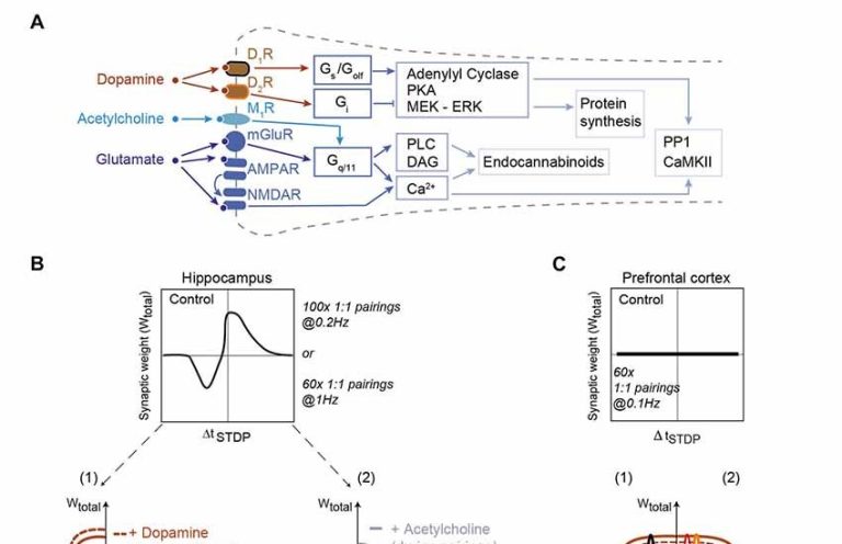
Written by Tjeerd V. olde Scheper et al. for Frontiers in Computational Neuroscience Vol. 11 - 2017 on January 10, 2018.
Spike Timing-Dependent Plasticity has been found to assume many different forms. The classic STDP curve, with one potentiating and one depressing window, is only one of many possible curves that describe synaptic learning using the STDP mechanism. It has been shown experimentally that STDP curves may contain multiple LTP and LTD windows of variable width, and even inverted windows.

Written by Jonathan C. Horton and Daniel L. Adams for Philos Trans R Soc Lond B Biol Sci. on April 29, 2005.
This year, the field of neuroscience celebrates the 50th anniversary of Mountcastle's discovery of the cortical column. In this review, we summarize half a century of research and come to the disappointing realization that the column may have no function. Originally, it was described as a discrete structure, spanning the layers of the somatosensory cortex, which contains cells responsive to only a single modality, such as deep joint receptors or cutaneous receptors.

Written by Jeff Hawkins, Subutai Ahmad, and Yuwei Cui for Frontiers in Neural Circuits on October 24, 2017.
Neocortical regions are organized into columns and layers. Connections between layers run mostly perpendicular to the surface suggesting a columnar functional organization. Some layers have long-range excitatory lateral connections suggesting interactions between columns.
We need your consent to load the translations
We use a third-party service to translate the website content that may collect data about your activity. Please review the details in the privacy policy and accept the service to view the translations.

